🎁 Exclusive Discount Just for You!
Today only: Get 30% OFF this course. Use code MYDEAL30 at checkout. Don’t miss out!
Also, you will be able to see subtle and high-quality details.-Risk signs on 12-Lead EKG These are concerns preoperatively, in ED or in clinic.
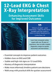
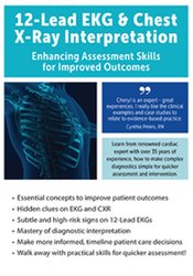
Cheryl Herrmann – 12-Lead EKG & Chest X-Ray Interpretation

PART I: THE ABCS of 12-LEAD EKG INTERPRETATION
12-Lead EKG 101
- Cardiac electric conduction system
- Electric vectors
- Introduction to the 12-Leads
- Limb leads
- Augmented leads
- Chest Leads
- Introduction to the 12-Leads
- Normal polarity, and P-QRS-Each lead has a T configuration
Ace the Axis
- Abnormalities in the left and right axes
- The causes and criteria for axis deviation
- Methods to determine axis deviation
- Clinical application, case analysis and presentation
Beat the Bundles
- Blocks of right-and left-bundled branch blocks (BBB).
- Criteria for right-vs.-left BBB
- BBB: Causes and complications
- Left anterior and left posterior haemiblocks
- Clinical application, case analysis and presentation
Correlate the Coronary Anatomy
- Coronary arteries
- Right coronary artery
- Left coronary artery (LCA).
- Left anterior descended artery (LAD).
- Circumflex artery
- Posterior descending artery (PDA).
- Left ventricular wall
- Inferior
- Anterior
- Septal
- Lateral
- Posterior
- Relationship between coronary arteries & ventricular walls to the 12-Leads
- Inferior—RCA—leads II, III, AVF
- Anterior/septal—LAD—leads V1–V4
- Lateral—circumflex—leads I, AVL, V5, V6
- Posterior—PDA—reciprocal changes V1–V4
Differential Diagnosis: 12-Lead EKG Acute Coronary Syndrome
- Ischemia pattern
- Injury pattern
- Infarction pattern
- Reciprocal Changes
- ST segment elevation myocardial infarction (STEMI)
- Non-ST segment elevation myocardial infarction (NSTEMI)
- Coronary spasm
- Takotsubo cardiomyopathy (broken heart syndrome)
Analyse and Example Time – Putting It All Together
- 30-On 12th, second diagnosis of STEMI-Lead EKG
- Correlation between Coronary angiographic and 12-Lead EKG
- Clinical application, case analysis and presentation
Advanced 12-Lead: Challenging Clinical Presentations
- Hypertrophy of the ventricular and atrial muscles
- Wolff-Parkinson-White syndrome
- Extended QT intervals
PART II: CHESTX-RAY INTERPRETATION-AS EASY AS BLACK & WHITE
Note: Most chest X-AP films will also be shown as ray examples
Chest X-Ray basics
- Technique
- Principles of black and white
- Projections
- Anterior-posterior (AP).
- Posterior-anterior (PA)
- Lateral
A Systematic Approach
- Bone structures
- Intercostal spaces
- Soft tissues
- Lungs/trachea/pulmonary vasculature
- Pleural surfaces
- Diaphragm
- Mediastinum
- Great vessels and the heart
- Invasive lines
As easy as Black
- Pneumothorax
- Subcutaneous emphysema
- Clinical application and treatment
It’s as easy as white
- Pleural effusion
- Pulmonary edema
- Pneumonia
- Atelectasis
- Acute respiratory distress syndrome (ARDS).
- Cardiomyopathy
- Pericardial effusion
- Cardiac Tamponade
- Clinical application and treatment
Beyond the Basics
- Aortic aneurysm
- Post-Op changes after pneumonectomy
- Hydropneumothorax
- Esophagogastrectomy
- Dextrocardia
- Application of clinical treatment
PART III: CXR & 12 LEAD EKG CASE PRESENTATIONS, CLINICAL APPLICATIONS AND ANALYSIS
Would you like a gift? Cheryl Herrmann – 12-Lead EKG & Chest X-Ray Interpretation ?
Description:
When to be concerned — 12-Lead EKG Pearls of Wisdom
When the coronary artery becomes blocked, the clock starts to tick. Are you able to interpret STEMI (ST-segment elevation myocardial injury) changes quickly using a 12-Lead EKG Provide effective intervention to reduce AMI size The 12-Lead EKG It is one of the most commonly used cardiac assessment tools to evaluate chest pain. It provides so much more information to improve patient care. This section of the recording is designed to help the health care professional master 12-Lead EKG Assessing skills to recognize the common and not-So-These are the most common changes. We will also look at subtle and high quality changes.-Risk signs on 12-Lead EKG These are concerns preoperatively, in ED or in clinic.
As Easy as Black and White…
Chest X-Radiation is also a diagnostic test that is commonly ordered, but it can be difficult to see and interpret them. Chest X-Rays can be as simple and straightforward as black or white. This portion of the recording provides clinicians with chest-X information.-ray interpretation skills in order to help you easily assess changes in the patient’s condition. You will learn how to identify lines, differentiate between pneumonia, atelectasis and pleural effusion and pulmonary edema, as well as identify cardiac tamponade and cardiomyopathy. There will be many examples of chest X.-to strengthen the concepts. Clinicians will leave this session with the practical skills to improve patient assessment and allow for more effective intervention.
Here’s what you’ll get in Cheryl Herrmann – 12-Lead EKG & Chest X-Ray Interpretation

Cheryl Herrmann – 12-Lead EKG & Chest X-Ray Interpretation Sample
Course Features
- Lectures 1
- Quizzes 0
- Duration Lifetime access
- Skill level All levels
- Language English
- Students 41
- Assessments Yes


